Solutions
keyboard_arrow_downServices
keyboard_arrow_downSupport & Resources
keyboard_arrow_downCompany
keyboard_arrow_downContact
Programmed Cell Death Protein 1 (PD-1) is a protein found on the surface of T cells, which are crucial for the immune system's ability to recognise and eliminate infected or damaged cells, including cancer cells.
PD-1 inhibits T cell activity to prevent excessive immune responses which can be damaging. A soluble form of PD-1 can be measured in the blood and is associated with various autoimmune diseases, cancers, and chronic infections.
Get in touch to discover more
To find out more about the Novel Biomarkers – PD-1, enquire now.
Autoimmune Diseases
Higher levels of sPD-1 have been linked to conditions like rheumatoid arthritis. T cells are key players in the development of rheumatoid arthritis, increased expression of sPD-1 may suppress PD-1 inhibition of T Cells, leading to prolonged T Cell activity and disease progression. Due to its role in regulating T cell activity, the PD-1 pathway is a potential target for rheumatoid arthritis therapy.
sPD-1 may also be involved in the immune dysfunction associated with other autoimmune diseases such as lupus, myasthenia gravis, and systemic sclerosis.
Other Applications
Cancer: sPD-1 levels are higher in cancers such as liver cancer, lung cancer, and pancreatic cancer. Monitoring sPD-1 can reflect tumour burden and help in evaluating treatment response. The PD-1 pathway is an attractive target for cancer treatment as blocking the interaction of PD-1 may stop PD-1 mediated inhibition of T Cell activation allowing T cells to target and destroy tumour cells.
Infection: Increased sPD-1 levels are found in chronic infections like hepatitis B and HIV, levels showed correlation with viral load.
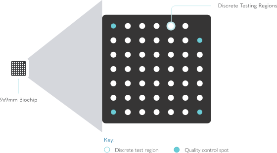
Biochip Array Technology
Randox biochip technology enables precise measurement of sPD-1, providing valuable insights into immune regulation and disease progression.
The Evidence MultiSTAT
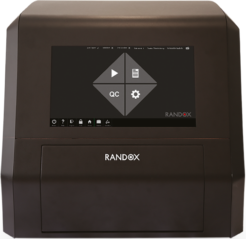
Evidence MultiSTAT
The Evidence MultiSTAT is an easy to use, small footprint analyser facilitating on-site simultaneous detection of multiple biomarkers.
Using chemiluminescence as a measurement principle, the Evidence MultiSTAT consistently delivers accurate results.
With minimal sample preparation required, this versatile benchtop analyser can achieve accurate, quantitative results in minutes.
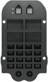
MultiSTAT Cartridge
The Evidence MultiSTAT cartridge contains the reagents required for the chemiluminescent reaction to take place incorporated into its wells.
The process from sample entry to results can be completed in 2 simple steps, with minimal risk of human error.
No other components are required.